Kaizen Fistula Care

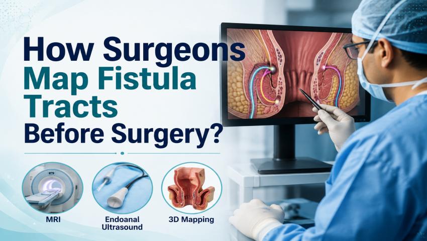
How Surgeons Map Fistula Tracts Before Surgery?

Fistula surgery is not simple. Every patient’s fistula is different. The shape, depth, and direction can all vary. A small mistake during surgery can harm the muscles. It can also leave a hidden part of the fistula untreated. Both problems lead to the same result — the fistula comes back. This is why mapping the fistula tract before surgery is so important.

At , Pune, Dr. Samrat Jankar follows a clear step-by-step method for fistula mapping. He uses his 17+ years of experience along with modern imaging tools. This helps him plan safe surgery, even in tricky and repeat cases. In this blog, we explain how surgeons map fistula tracts, which tests are used, and why this step is so important for successful treatment.

What Is an Anal Fistula?

An anal fistula is like a small tunnel. It forms between the inside of the anal canal and the skin near the anus. Most fistulas start after an anal abscess does not heal fully. A small channel is left behind, and infection keeps coming out from it.

Common signs of fistula are:

  • Regular discharge of pus or blood near the anus
  • Pain and swelling, more while sitting
  • Skin irritation around the anus
  • Fever during flare-ups
  • A small opening on the skin that keeps leaking

If not treated, an anal fistula does not heal on its own. It keeps causing infection and discomfort. This is why early check-up and correct tract mapping are so important before starting treatment.

Why Is Fistula Tract Mapping Done Before Surgery?

Each fistula tract behaves differently inside the body. Some tracts are short and straight. Others turn, branch out, or connect to more than one opening on the skin. Without knowing the full path, the surgeon cannot plan well.

Fistula tract mapping helps the surgeon to:

  • See the full length and direction of the tract
  • Check how close the tract is to the anal sphincter muscles
  • Find hidden branches or pus pockets
  • Know if the fistula is simple or complex
  • Plan the surgery in a way that protects bowel control

Good mapping does two important things. It lowers the chance of the fistula coming back. It also protects the patient’s ability to control bowel movements after surgery.

How Do Surgeons Identify the Fistula Tract?

Surgeons never depend on just one method. They use a mix of physical check-up and imaging tests together. This gives a full and correct picture of the fistula before surgery. Skipping this step is one of the biggest reasons fistulas come back after poor planning.

Initial Examination Before Fistula Surgery:

  • Medical History and Symptoms Assessment: The first step is always a detailed talk with the patient. The doctor asks how long the symptoms have been there. He also asks about the type of discharge, pain level, and any fever or past abscess. Old treatments, past fistula surgeries, and health issues like diabetes are also noted. This helps the doctor understand the likely cause of the fistula.
  • Physical Examination of the Fistula: Next, the doctor does a hands-on check. He finds the outer opening on the skin. He checks for discharge, pain, redness, and swelling. A gentle rectal examination is often done to feel the inner opening. This gives early clues about the direction of the tract, even before any test.

Tests Used to Map Fistula Tracts Before Surgery:

  • MRI Scan for Anal Fistula: MRI, or Magnetic Resonance Imaging, is known as the best test for fistula mapping. It uses magnetic fields to give clear pictures of soft tissue. There is no radiation used. MRI clearly shows the full path of the tract, any side branches, hidden pus, and how close it is to the sphincter muscles. For complex or repeat fistulas, MRI is almost always advised.
  • Endoanal Ultrasound (EAUS): This test uses a small ultrasound probe placed gently inside the anal canal. Sound waves create live images of the muscles and nearby tissue. EAUS is quick and fairly comfortable. It works well for simple, low fistulas. It also helps check the health of the sphincter muscles.
  • CT Scan (When Is It Required?): A CT scan is not used often for simple fistula cases. It is useful when there is a deep infection, or when the abscess is spreading towards the pelvis or other organs. In such cases, CT gives a wider view beyond just the fistula tract.
  • Examination Under Anaesthesia (EUA): Sometimes, even the best scans cannot fully explain a tricky tract. In such cases, the surgeon checks the area under anaesthesia, just before or at the start of surgery. This gives a clear, hands-on check using probes and dye. The patient feels no pain, as this is done under sedation.

How MRI Helps Surgeons Plan Fistula Surgery?

MRI plays a big role in planning the surgery. It helps in many ways:

  • Shows the exact path of the tract — including turns, branches, or closed pockets that a physical check may miss.
  • Shows how close the tract is to the sphincter muscles — this is important, because cutting too much muscle can affect bowel control for life.
  • Finds hidden infection or pus — this needs to be drained before or during the main surgery.
  • Helps classify the fistula correctly — which decides the best surgery method.

With this information, the surgeon can enter the operation theatre with almost a full map. This lowers the chance of surprises during surgery.

Different Types of Fistula Tracts Surgeons Look For:

Fistulas are grouped based on how the tract relates to the sphincter muscles. This grouping helps decide the right surgery.

  • Intersphincteric Fistula This tract stays between the inner and outer sphincter muscles. It does not cross the outer muscle. It is the most common type. It is usually easier to treat, with a good success rate.
  • Transsphincteric Fistula This tract crosses both the inner and outer sphincter muscles before opening on the skin. Since it passes through more muscle, it needs careful planning. This protects long-term bowel control.
  • Suprasphincteric Fistula This tract goes above the sphincter muscles, loops over them, and then comes down to the skin. It is more complex than the first two types. It often needs a mixed treatment approach.
  • Extrasphincteric Fistula This is a rare type. It fully bypasses the sphincter muscles. It usually comes from another cause, like a pelvic or bowel problem. It often needs care from more than one specialist.
  • Complex and Recurrent Fistulas Some fistulas have many openings or branching tracts. Some come back after a past surgery done elsewhere. These cases need detailed imaging, careful mapping, and strong surgical skill for a lasting cure.

How Fistula Mapping Helps in Choosing the Right Surgery?

Once the tract is mapped correctly, the surgeon can choose the best treatment. The choice depends on the type, depth, and complexity of the fistula.

  • Fistulotomy: The full tract is opened up. This works well for simple, low fistulas that do not involve much muscle.
  • Seton Placement: A soft thread, called a seton, is passed through the tract. This allows slow drainage and healing. It is useful for complex tracts where quick cutting can risk bowel control.
  • LIFT Procedure (Ligation of Intersphincteric Fistula Tract): This method closes the tract from between the sphincter muscles, without cutting the muscle. It helps mainly in transsphincteric fistulas.
  • Advancement Flap Surgery: Healthy tissue is used to cover the inner opening of the fistula. This is often chosen for high or complex tracts, to save the muscle.
  • Laser Treatment for Fistula: This is a minimally invasive option. It uses laser energy to seal the tract from inside. It usually gives a shorter recovery time and less pain after surgery.

Without proper mapping, the surgeon may pick the wrong method. This raises the risk of the fistula coming back. Correct mapping makes sure the right surgery is chosen the first time. This saves the patient from repeat surgeries.

Common Challenges While Mapping Complex Fistulas:

Not every fistula is easy to map. Some tracts curve sharply. Some branch into two or three directions. Some hide small pus pockets that are easy to miss. Old scar tissue from a past surgery can also make the images harder to read. Repeat fistulas are especially tricky, since the earlier surgery has already changed the natural shape.

This is where experience really matters. A skilled Fistula Specialist in Pune, like Dr. Samrat Jankar, brings years of hands-on surgery experience to read these results correctly. His work on thousands of fistula surgeries, along with his own PILTEC technique for complex and repeat cases, helps him plan surgery with much more confidence and accuracy.

What Patients Should Expect Before Fistula Mapping?

Knowing what happens before your check-up can reduce a lot of worry.

  • You may be asked to fast for a few hours before an MRI scan.
  • The MRI scan usually takes 30 to 45 minutes. It is completely painless.
  • No injections or cuts are usually needed for a normal MRI or ultrasound.
  • The doctor will look at the images himself and explain the results in simple language.
  • Based on the mapping results, the treatment plan and the surgery option will be discussed with you clearly, along with the expected recovery time.

There is no need to feel nervous about these tests. They are done to make your surgery safer and more predictable, not to add stress.

Benefits of Accurate Fistula Tract Mapping:

  • Greatly reduces the chance of the fistula coming back after surgery
  • Protects the sphincter muscles and keeps bowel control normal
  • Helps the surgical team pick the correct, most effective method
  • Improves the overall success rate of treatment, especially in tough cases
  • Gives the surgeon a clear plan before surgery, so there are fewer surprises
  • Builds patient trust, since the treatment plan is based on solid test results, not guesswork

Tips to Prepare for Your Fistula Assessment:

  • Share your full medical history with the doctor, including any past abscess or fistula treatment.
  • Tell the doctor about any long-term issues like diabetes, Crohn’s disease, or past pelvic surgery.
  • Follow the fasting instructions given before your MRI scan carefully.
  • Wear loose, comfortable clothes on the day of your scan.
  • Keep a note of your symptoms, including when they started and how they have changed.
  • Bring any old scan reports or surgery records to your visit, if you have them.
  • Do not be shy to ask the doctor questions about the results or the planned treatment.

Conclusion:

Mapping the fistula tract is the real base of successful fistula surgery. It helps the surgeon know the exact path and type of the tract before making even one cut. This lowers the surgical risk, protects bowel control, and improves healing.

At Kaizen Fistula Care, Pune, Dr. Samrat Jankar brings 17+ years of surgical experience and modern imaging tools to plan safe, effective treatment. If you are looking for a trusted Fistula Specialist in Pune, remember — correct mapping is the first step towards full relief.

Do not ignore fistula symptoms. Book your appointment with Dr. Samrat Jankar today.

Call 79041 39064 or email kaizenfistulacare@gmail.com to book. Visit us at Kaizen Gastro Care, Pune, Maharashtra 411057.

Frequently Asked Questions:

MRI is not needed for every case, especially very simple, low fistulas that are clear on a physical check. But for complex, repeat, or high fistulas, MRI is strongly advised. It gives the most correct and detailed picture of the tract, its branches, and its closeness to the sphincter muscles.

No, fistula mapping is usually not painful. Tests like MRI and endoanal ultrasound do not need any cuts and cause little to no discomfort. If an examination under anaesthesia is needed, it is done under sedation, so the patient feels no pain.

MRI is seen as the best and most reliable test for detailed fistula mapping, especially for complex or repeat cases. Endoanal ultrasound is a good option for simpler fistulas and is quicker to do, but MRI usually gives more detail for surgery planning.

MRI finds most fistula tracts correctly, including hidden branches and pus pockets. But in a few very complex or heavily scarred cases, an examination under anaesthesia may still be needed at the time of surgery, for full confirmation.

Yes, definitely. Correct mapping lets the surgeon pick the right surgery method for the exact type of fistula. It also helps avoid leaving any hidden branch or infected tissue untreated. Both these things lower the risk of the fistula coming back after surgery.